Dermoscopic features of amelanotic and hypomelanotic melanoma
Melanoma
Skin cancer
Case presentations
Case 1
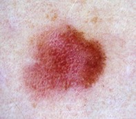
A 34-year-old woman was referred by her GP for review of an asymptomatic pink lesion on her thigh. The main concern was that the lesion had grown slightly. She had no history of significant sun exposure. Clinical examination revealed a slightly elevated pink plaque (7 x 4 mm), which appeared relatively symmetrical (Figure 1a).
On dermoscopy, the lesion was a homogeneous pink colour with a central white area, most probably caused by the pressure of the non-polarised contact dermatoscope. Numerous dotted and linear irregular vessels were visible throughout the lesion (Figure 1b).
The lesion was excised with a 2 mm margin. Histopathological examination showed a 0.3 mm level II melanoma.
Single article purchases are temporarily unavailable due to site maintenance.
If you would like to purchase an article during this time, please email us at [email protected] with the article details and we'll assist you directly. We'll also let you know when online purchasing is available again.
Thank you for your patience and understanding.