Beware blind spots!
Skin lesions
Skin cancer
Case presentations
Case 1
A 68-year-old woman presented for assessment of two skin lesions, which were diagnosed as a solar keratosis on the left cheek and a superficial basal cell carcinoma (BCC) on the presternal chest. After discussing treatment options with respect to her presenting complaints, a full skin examination was performed.
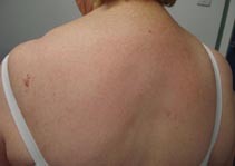
On initial inspection, the patient’s back appeared to be clear of any lesions of concern (Figure 1a). After displacing the bra-straps, a prominent vascular lesion measuring over 2 cm in maximum diameter was noted over the left upper back (Figures 1b and c). On dermoscopy, multiple cherry-red dots, globules and lacunes were observed (Figure 1d). When questioned further, the patient mentioned that the lesion was longstanding.
The provisional clinical diagnosis was angioma serpiginosum. A biopsy was not considered necessary. The patient was reassured that the lesion was benign and not requiring treatment.
Single article purchases are temporarily unavailable due to site maintenance.
If you would like to purchase an article during this time, please email us at [email protected] with the article details and we'll assist you directly. We'll also let you know when online purchasing is available again.
Thank you for your patience and understanding.